

External Cervical Resorption: Which lesions are treatable?
July 1, 2026
By Lisa Germain, DDS, MScD
Introduction
External cervical resorption (ECR) is one of the more challenging forms of tooth resorption encountered in clinical practice. It begins on the external surface of the tooth near the cemento-enamel junction and can progress silently for years before being detected. In many cases, patients experience no symptoms until significant tooth structure has already been lost.
Advances in cone-beam computed tomography (CBCT) have improved our ability to diagnose external cervical resorption, but determining whether a tooth can be saved still depends largely on understanding the extent of the lesion. One of the most widely recognized systems for evaluating ECR was developed by Dr. George Heithersay, who divided lesions into four classifications based on their size and depth of invasion.
Understanding these classifications is important because treatment options and long-term prognosis vary considerably from one class to another. Small lesions can often be treated predictably, while more advanced lesions may ultimately require extraction.
What Is External Cervical Resorption?
External cervical resorption is a condition in which specialized cells begin to resorb the cementum and dentin of a tooth. Unlike dental caries, which result from bacterial activity, ECR is caused by clastic cells that destroy tooth structure from the outside inward.
The condition most commonly develops in the cervical region of the tooth, just below the gumline. Although the exact cause is not always known, several factors have been associated with an increased risk of ECR, including:
- Orthodontic treatment
- Dental trauma
- Internal bleaching procedures
- Periodontal therapy
- Surgical procedures involving the teeth
- Bruxism and occlusal trauma
- Idiopathic factors
One of the unique features of ECR is that the pulp often remains healthy despite extensive destruction of surrounding dentin. As a result, patients frequently report no symptoms until the lesion is discovered during a routine examination or radiographic evaluation.
In 1999 GS Heithersay created a classification system for ECR based on the location and size of the lesion. In doing so, he clarified the parameters in which it can be determined whether the lesions are predictably treatable. A diagram is provided below.
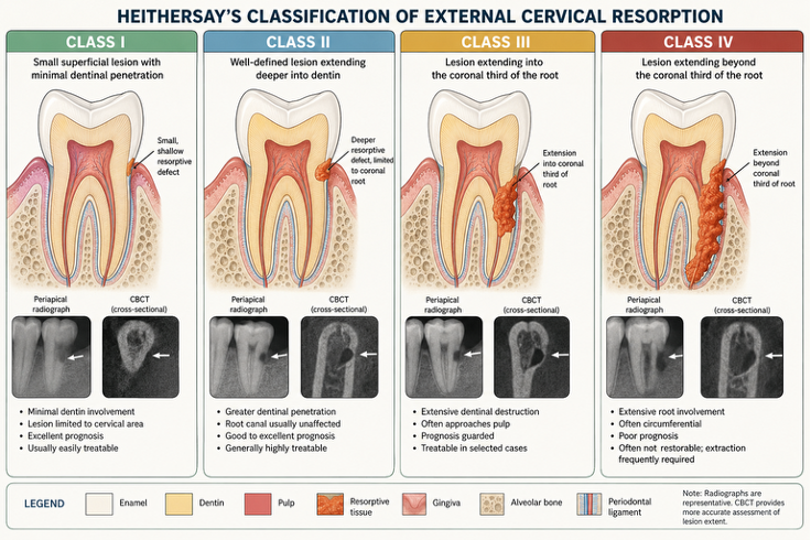
Heithersay Class I
Class I lesions represent the earliest and least destructive form of external cervical resorption. The lesion is small and superficial, with only minor penetration into dentin.
Clinically, these lesions may appear as a small pink discoloration near the cervical area of the tooth or may be detected incidentally on radiographs. Because the lesion is localized and easily accessible, treatment is usually straightforward.
After gaining access to the defect, the clinician removes all resorptive tissue and restores the area with a suitable restorative material. Root canal treatment is rarely necessary unless the pulp becomes exposed during treatment.
The prognosis for Class I lesions is excellent, and long-term retention of the tooth is highly predictable.
Heithersay Class II
Class II lesions extend deeper into dentin but remain relatively localized. Although the lesion is larger than a Class I defect, it generally has not spread significantly into the root structure.
Most Class II lesions can still be treated successfully. Treatment typically involves surgical access, removal of all resorptive tissue, and restoration of the defect. Depending on the lesion’s proximity to the pulp, endodontic therapy may occasionally be required.
When diagnosed early and managed appropriately, Class II lesions generally have a very favorable prognosis.
Heithersay Class III
Class III lesions represent a more advanced stage of external cervical resorption. The defect extends deeper into the tooth and often reaches the coronal third of the root.
At this stage, treatment becomes more complex. Conventional radiographs frequently underestimate the true size of the lesion, making CBCT imaging an essential part of diagnosis and treatment planning.
Management usually requires surgical exposure, complete debridement of the resorptive tissue, and restoration of the defect. Because of the lesion’s depth, root canal therapy is often necessary either before or during treatment.
The prognosis for Class III lesions is considered guarded. Success depends on the ability to completely access the lesion and preserve sufficient tooth structure for long-term function.
Heithersay Class IV
Class IV lesions are the most extensive form of external cervical resorption. The lesion extends beyond the coronal third of the root and may involve a large portion of the root circumference.
These lesions present significant treatment challenges. In many cases, extensive destruction of dentin leaves insufficient tooth structure to support a predictable restoration. Even when treatment is technically possible, the risk of recurrence and structural failure is substantially higher.
For this reason, extraction is often the most appropriate treatment option for Class IV lesions. Replacement options may include dental implants, fixed partial dentures, or removable prostheses depending on the individual patient’s circumstances.
How Are Treatable Lesions Managed?
The specific treatment approach varies depending on lesion size and location, but most successful ECR treatment follows several common steps:
- Diagnosis with clinical examination and CBCT imaging.
- Surgical access to the lesion.
- Removal of all resorptive tissue.
- Restoration of the defect using biocompatible materials such as composite resin, Biodentine, or mineral trioxide aggregate (MTA).
- Endodontic treatment when pulpal involvement is present.
- Periodic clinical and radiographic follow-up.
The primary objective is to completely eliminate active resorptive tissue while preserving as much healthy tooth structure as possible.
Which Lesions Can Be Saved?
As a general guideline:
- Class I lesions are almost always treatable.
- Class II lesions are usually treatable with predictable outcomes.
- Class III lesions are often treatable but require careful case selection.
- Class IV lesions frequently have a poor prognosis and may not be restorable.
The earlier external cervical resorption is detected, the greater the likelihood of successful treatment and long-term tooth retention.
Conclusion
Heithersay’s classification system remains one of the most useful tools for evaluating external cervical resorption and determining treatment prognosis. While Classes I and II can often be managed successfully with conservative intervention, Classes III and IV require increasingly complex treatment and carry a less favorable prognosis. Modern CBCT imaging has greatly improved clinicians’ ability to diagnose these lesions accurately, allowing for better treatment planning and more predictable outcomes.
Ultimately, early diagnosis remains the key factor in preserving teeth affected by external cervical resorption.
Bibliography
-
- Heithersay GS. “Invasive Cervical Resorption Following Trauma.” Australian Endodontic Journal. 1999;25(2):79–85.
- Heithersay GS. “Clinical, Radiologic, and Histopathologic Features of Invasive Cervical Resorption.” Quintessence International. 1999;30(1):27–37.
- Heithersay GS. “Invasive Cervical Resorption: An Analysis of Potential Predisposing Factors.” Quintessence International. 1999;30(2):83–95.
- Patel S, Kanagasingam S, Pitt Ford T. “External Cervical Resorption: A Review.” Journal of Endodontics. 2009;35(5):616–625.
- Patel S, Foschi F, Mannocci F, Patel K. “External Cervical Resorption: A Three-Dimensional Classification.” International Endodontic Journal. 2018;51(2):206–214.
- Tronstad L. Clinical Endodontics. 3rd ed. New York: Thieme; 2009.
- Andreasen JO, Andreasen FM, Andersson L. Textbook and Color Atlas of Traumatic Injuries to the Teeth. 5th ed. Oxford: Wiley-Blackwell; 2018.
- Fuss Z, Tsesis I, Lin S. “Root Resorption—Diagnosis, Classification and Treatment Choices Based on Stimulation Factors.” Dental Traumatology. 2003;19(4):175–182.
- Bergmans L, Van Cleynenbreugel J, Verbeken E, Wevers M, Van Meerbeek B, Lambrechts P. “Cervical External Root Resorption in Vital Teeth.” Journal of Clinical Periodontology. 2002;29(6):580–585.
- Patel S, Saberi N. “The Ins and Outs of Root Resorption.” British Dental Journal. 2018;224(9):691–699.