Maxillary Sinusitis of Endodontic Origin
January 1, 2024
By Lisa Germain, DDS, MScD
Odontogenic sinusitis is a broad term used to describe any degree of sinus infection and symptoms that are caused by dental pathology. Among the various multiple dental etiologies are periodontal disease, endodontic disease, root fractures, dental implants, dental extractions, oral-antral fistulae, and iatrogenic causes such as extruded dental materials, displaced teeth, and foreign bodies. (1-9) While these can all be odontogenic sources for sinusitis, it is important to distinguish these etiologies from maxillary sinusitis of endodontic origin (MSEO), as they each have a different pathogenesis and require markedly different clinical treatments. MSEO refers specifically to sinusitis caused by endodontic infection, excluding sinusitis secondary to other dental etiologies. Another way to categorize this is to say that there is primary endodontic pathosis which is causing secondary sinus pathosis. Often when the primary source is eliminated, the secondary source resolves without further treatment.
Maxillary sinusitis of endodontic origin (MSEO) was first referred to in the dental literature in 1943. This phenomenon is seen in 60-80% of patients with infections originating in the maxillary posterior teeth and is the most common form of odontogenic sinusitis. The literature also indicates that dental infections may account for more than 40% of maxillary sinusitis cases. (10-12)
Treating the primary source of any disease is of paramount importance. Hence, failure to recognize MSEO and properly manage the endodontic pathosis will result in the persistence of sinus disease and the failure of medical sinus therapies. If left undiagnosed, patients often suffer with chronic sinus infections, ineffectual antibiotic regimens, and may even undergo multiple sinus surgeries, never realizing that an endodontic infection is the source. MSEO also has the potential to advance to more serious or even life-threatening cranio-facial infections. In these severe and rare cases, endodontic infection can spread via the maxillary sinus causing orbital cellulitis, blindness, meningitis, subdural empyema, brain abscess and life-threatening cavernous sinus thrombosis (13-17).
Patients with MSEO experience a wide variation of dental and sinonasal symptoms including no symptoms and diagnosing MSEO can be challenging since the history of the reported illness lacks the classic pattern usually associated with apical periodontitis commonly seen in teeth with a necrotic pulp. These teeth rarely have thermal symptoms, and most likely are not percussion sensitive because the abscess is draining into the sinus or displacing the floor of the sinus thus relieving the pressure(Fig 1). For this same reason, swelling or intraoral sinus tracts rarely form.

Figure 1: MSEO caused by necrotic pulp in tooth #14 with drainage into sinus.
Patients with MSEO are more likely to experience common sinonasal symptoms, which include congestion, runny nose, facial pain, and a foul odor. (18, 19) Patients with sinonasal symptoms that present without localized dental pain will often first seek care from their primary care physician or ENT specialist who may misdiagnose and treat MSEO as a primary sinus infection since a dental source is not tested for during routine ENT examinations. Current ENT clinical guidelines for the medical management of rhinosinusitis offer no guidance in odontogenic etiologies and there is no mention of dental infections as a potential cause of sinusitis. (19) For physicians and ENT specialists, findings that should raise the suspicion of MSEO are a history of repeated episodes of unilateral maxillary sinus infections, particularly when associated with a patent sinus ostium or previously unsuccessful sinus surgery. (Fig 2) (20) While the diagnosis of sinonasal disease secondary to endodontic infection is within the scope of dentistry since treatment of the tooth is indicated, dentists. should not attempt to make a final diagnosis of non-odontogenic sinus disease (primary sinusitis), nor offer treatment that is outside the scope of dental practice.

Figure 2: Sinus pathosis is unilateral which is frequently seen in cases with MSEO.
Historically, periapical radiographs are the most widely used imaging modality in endodontics, the posterior maxilla presents significant and unique interpretation challenges when using conventional 2D imaging. Anatomic structures such as the zygoma, palatal process, maxillary sinus, and buccal cortical plate are often superimposed over the dental roots, obscuring or concealing peri-radicular infection (Fig 3a). Conventional periapical radiographs also do not consistently reveal mucosal thickening or fluid in sinuses, which are of important diagnostic value in MSEO. CBCT imaging has been shown to significantly improve the ability to detect odontogenic sources for sinusitis. (21) In a study by Low et al. (22), CBCT revealed 34% more lesions than periapical radiography, as well as significantly more expansion of lesions into the maxillary sinus, mucosal thickening, and untreated canals. Mucosal changes associated with dental infections were found with a prevalence of 77%, compared to only 19% using conventional radiographs. (Fig.3b)
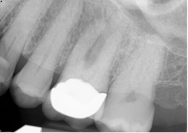
Figure 3a: 2-dimensional periapical radiograph of upper molar (#14) with superimposed anatomical structures concealing the severity of the pathology.

Figure 3b: CBCT shows definitive peri-radicular pathology and displacement into sinus floor due to swelling caused by pulp necrosis of upper molar (#14) shown in 2-dimensional image in figure 3a.

Figure 4: CBCT image of necrotic tooth #14 draining into sinus with evidence of mucosal thickening.
A thorough clinical endodontic examination is essential for diagnosing or ruling out MSEO. When diagnosing a possible endodontic etiology in patients with sinusitis, the clinician must look carefully for any teeth with pulpal necrosis and evaluate all prior endodontic treatments for possible failure in the suspected quadrant. Because MSEO is a bacterial disease, typically, only teeth with an infected necrotic pulp or failing endodontic treatment will cause significant sinonasal disease or sinonasal symptoms. When examining maxillary posterior teeth with existing root canal treatment, one must carefully examine for any untreated or sub-optimally filled canals, inadequate core restorations, or leaking coronal restorations that may provide evidence of endodontic failure and a bacterial source for MSEO.
The objectives for treatment of MSEO are removal of the pathogenic microorganisms, their by-products, and pulpal debris from the infected root canal system that are causing the sinus infection and preventing reinfection. Appropriate treatment options include non-surgical root canal therapy, peri-radicular surgery when indicated, intentional replantation, or extraction of the infected tooth. Patients should be informed of all treatment options, the prognosis of each option and the risks of no treatment. Maxillary molars typically have complex anatomy and inadequate root canal treatment, particularly missed mesio-buccal canal systems, is a common cause of endodontic failure in maxillary molars. The close anatomic proximity of maxillary molar root apices to the floor of the maxillary sinus can lead to persistent MSEO if canals are left untreated or root canal failure occurs in these teeth.
Use of systemic antibiotics to manage MSEO should follow the guidelines set forth in the AAE Guidance on the Use of Systemic Antibiotics in Endodontics. (23) Apart from spreading infections, antibiotic therapy is unwarranted in the treatment of MSEO and ineffective as a definitive solution. While antibiotic therapy may offer temporary relief of symptoms by improving sinus clearing, their sole use is inappropriate without definitive debridement and disinfection of the root canal system. Antibiotics are not effective in eliminating the infection contained within the root structure and can only be eliminated by cleaning, shaping, sterilizing and obturating the root canal system. Similarly, surgical intervention of the maxillary sinus that is focused strictly on removing diseased sinus tissue, and establishing drainage is inadequate if the endodontic component is not eliminated.
The dental literature provides numerous case reports showing full resolution of MSEO following endodontic treatment. (24-30) It should be noted, however, that endodontic treatment alone may not resolve all cases of MSEO, therefore clinical and radiological follow-up is essential as concomitant management of the associated rhinosinusitis by an ENT specialist may be necessary in some cases. (31-33) A collaborative effort and open referral relationships between general dentists, endodontists, and ENT surgeons is essential to achieve the best outcomes for patients with MSEO.
Conclusion
MSEO is fundamentally an endodontic infection manifesting in the maxillary sinus and is a common, yet often overlooked disease process. Symptoms and radiographic signs of MSEO often mimic primary sinusitis leading patients to first seek care from their primary care physician or ENT specialist, whose treatment will not resolve MSEO if the endodontic source is not addressed. MSEO is also frequently overlooked in general dental practice due to a lack of classic dental symptoms and an obscured or atypical radiographic presentation. In-office cone-beam computed tomography has increased clinicians’ recognition and ability to diagnose MSEO. Clinical endodontic examination, however, remains essential for correct diagnosis that manifests in the maxillary sinus. Solid referral relationships and improved communication between general dentists, endodontic specialists and ENT surgeons are critical to providing appropriate patient care when managing MSEO.
References
- Abrahams JJ, Glassberg RM. Dental disease: a frequently unrecognized cause of maxillary sinus abnormalities? Am J Roentenol 1996;166:1219-23.
- Mehra P, Murad H. Maxillary sinus disease of odontogenic origin. Otolarngol Clin North Am 2004;37:347-64.
- Kretzschmar DP, Kretzschmar JL. Rhinosinusitis: Review from a dental perspective. Oral Surg Oral Med Oral Pathol Oral Radiol Endod 2003;96:128-35.
- Legert KG, Zimmerman M, Stierna P. Sinusitis of odontogenic origin: pathophysiological implications of early treatment. Acta Otolaryngol 2004;124:655-63.
- De Lima CO, Devito KL, Vasconselos LR, et al. Correlation between endodontic infection and periodontal disease and their association with chronicsinusitis: A clinical- tomographic study. J Endod 2017;43:1978-83.]
- Zirk M, Dreiseidler T, Pohl M, et al. Odontogenic sinusitis maxillaris: A retrospective study of 121 cases with surgical intervention. J Craniomaxillofac Surg
- Lechian JR, Filleul O, Costa de Araujo P, et al. Chronic maxillary rhinosinusitis of dental origin: A systematic review of 674 patient cases. Int J Otolaryngol
- Taschieri S, Torretta S, Corbella S, et al. Pathophysiology of sinusitis of odontogenic origin. J Investig Clin Dent 2017;8(2).
- Maillet M, Bowles WR, McClanahan, SL. Cone-Beam computed tomography evaluation of maxillary sinusitis. J Endod 2011;37:753-57.
- Bomelli SR, Branstetter BF, Ferguson, BF. Frequency of a dental source for acute maxillary sinusitis, Laryngoscope 2009; 119(3):580-84.
- Matsumoto Y, Ikeda T, Yokoi H, et al. Association between odontogenic infections and unilateral sinus opacification. Auris Nasus Larynx 2015;42:288-93.
- Patel NA, Ferguson BJ. Odontogenic sinusitis: an ancient but under-appreciated cause of maxillary sinusitis. Curr Opin Otoloaryngol Head Neck Surg 2012;20:24-8.
- Obayashi, N., Ariji, Y., Goto, M. et al. Spread of odontogenic infection originating in the maxillary teeth: computerized tomographic assessment. Oral Surg Oral Med Oral Pathol Oral Radiol Endod 2004;98:223–31.
- Eufinger H, Machtens E. Purulent pansinusitis, orbital cellulitis and rhinogenic intracranial complications. J Cranio Maxillofac Surg 2001;29:111-7.
- Wagenmann M, Naclerio RM. Complications of sinusitis. J Allergy Clin Immunol 1992;90:552-4.
- Gold RS, Sager E. Pansinusitis, orbital cellulitis, and blindness as sequelae of delayed treatment of dental abscess. J Oral Surg 1974;32:40-3.
- Park CH, Jee DH, La TY. A case of odontogenic orbital cellulitis causing blindness and severe tension orbit. J Korean Med Sci 2013;28:340-3.
- Workman AD, Granquist EJ, Adappa ND. Odontogenic sinusitis: developments in diagnosis, microbiology, and treatment. Curr Opin Otolaryngol Head Neck Surg 2018;26:27-33.
- Rosenfeld RM, Piccirillo JF, Chandrasekhar SS, et al. Clinical practice guideline (update): Adult sinusitis executive summary. Otolaryngol Head Neck Surg 2015;152:598–609.
- Pokorny A, Tataryn R. Clinical and radiologic findings in a case series of maxillary sinusitis of dental origin. Int Forum Allergy Rhinol 2013;3:973-9.
- Lofthag-Hansen S, Huumonen S, Grondahl K, et al. Limited cone-beam CT and intraoral radiography for the diagnosis of periapical pathology. Oral Surg Oral Med Oral Pathol Oral Radiol Endod 2007;103:114-9.
- Low, K.M., Dula, K., Burgin, W., and von Arx, T. Comparison of periapical radiography and limited cone-beam tomography in posterior maxillary teeth referred for apical surgery. J Endod 2008;34:557–62.
- Fouad AF, Byrne BE, Diogenes AR, et al. AAE Guidance on the Use of Systemic Antibiotics in Endodontics. Association of Endodontists Position Statement 2017:1-8.
- Nenzen B, Welander U. The effect of conservative root canal therapy on local mucosal hyperplasia in the maxillary sinus. Odontol Revy 1967;18:295-302.
- Dodd RB, Dodds RN, Hocomb JB. An endodontically induced maxillary sinusitis. J Endod 1984;10:504-6.
- Bogaerts P, Hanssens JF, Siquet JP. Healing of maxillary sinusitis of odontogenic origin following conservative endodontic retreatment: case reports. Acta Otorhinolaryngol Belg 2003;57:91-7.
- Wang KL, Nichols BG, Poetker DM, Loehrl TA. Odontogenic sinusitis: a case series studying diagnosis and management. Int Forum Allergy Rhinol 2015;5:597-601.
- Tomomatsu N, Uzawa N, Aragaki T, Harada K. Aperture width of the osteomeatal complex as a predictor of successful treatment of odontogenic maxillary sinusitis. Int J Oral Maxillofac Surg 2014;43:1386-90.
- Bendyk-Szeffer M, Lagocka R, Trusewicz M, et al. Perforating internal root resorption repaired with mineral trioxide aggregate caused complete resolution of odontogenic sinus mucositis: A case report. J Endod 2015;41:274-8.
- Nurbakhsh B, Friedman S, Kulkarni GV, et al. Resolution of maxillary sinus mucositis after endodontic treatment of maxillary teeth with apical periodontitis: A cone-beam computed tomographic pilot study. J Endod 2011;37:1504-11.
- Brook I. Microbiology of acute and chronic maxillary sinusitis associated with an odontogenic origin. Laryngoscope 2005;115:823-5.
- Simuntis R, Kubilius R, Vaitkus S. Odontogenic maxillary sinusitis: a review. Stomatologija 2014;16:39-43.
- Saibene AM, Pipolo GC, Lozza P, et al. Redefining boundaries in odontogenic sinusitis: a retrospective evaluation of extramaxillary involvement in 315 patients. Int Forum Allergy Rhinol 2014;4:1020-3.
- Article adapted from Colleagues for Excellence: Maxillary Sinusitis of Endodontic Origin, Fall 2018